Home
/ Tendon Diagram Under Microscope, Histological sample Elastic cartilage Tisue under the ... - Cross section human tendon under microscope view for education histology, human tissue, dense regular connective tissue.
Tendon Diagram Under Microscope, Histological sample Elastic cartilage Tisue under the ... - Cross section human tendon under microscope view for education histology, human tissue, dense regular connective tissue.
Tendon Diagram Under Microscope, Histological sample Elastic cartilage Tisue under the ... - Cross section human tendon under microscope view for education histology, human tissue, dense regular connective tissue.. Viewing hair under the microscope students can observe and study the characteristics of a hair fiber/strand including pigmentation, scales as well as the pattern of the medulla. Here's a diagram of a plant cell: Coloured scanning electron micrograph (sem) of tendon fibres. Here's a photo of a plant cell under an electron microscope. Human skin under microscope 400x.
Cell membrane dr jastrow s electron microscopic atlas. However, tendon cell activity during growth and homeostatic maintenance is less well defined. Cross section human tendon under microscope view for education histology, human tissue, dense regular connective tissue. Images of individual cells were captured at 0% strain as well as sequentially at 2%, 4% and 6. Tendons generally have a very complex structure;
Monocot Plant Vascular Tissue Under The Microscope View ... from media.istockphoto.com Tendons transmit skeletal muscle forces to bone and are essential in all voluntary movement. Tendons have a hierarchical arrangement that is sequentially composed of collagen molecules, fibrils, fibres, fascicles. Some of the fibres have been teased apart. Otherwise, all tendons would weaken and rupture (ker, 2002). Tendons and muscles work together to move bones. Learn vocabulary, terms and more with flashcards, games and other study tools. At the chair of medical biophysics the scientists also deployed micro computer tomography to represent the interface region in three dimensions. Sp8 lightning confocal microscope products leica microsystems.
Tendon is a relatively simple tissue, with one predominant cell type—fibroblasts, which in tendon are called tenocytes and which are embedded in an insoluble matrix of elongated collagen fibrils that are surrounded by a soluble compartment of glycoproteins including proteoglycans.
But at the same time it is interpretive. Tendons and ligaments containing progenitor cells. They are actually heavily composed of connective. In turn, movement appears to affect tendon properties, and. Apart from macroscopic investigations, the microscopic investigation of hair is a big part of forensic investigations. Transmission electron microscopy (tem) is a microscopy technique in which a beam of electrons is transmitted through a specimen to form an image. Tenocytes constantly repair small amounts of damage to the matrix under normal circumstances; Microscope information, images from beneath the microscope and educational science projects. Cell membrane dr jastrow s electron microscopic atlas. The tendon cells of the rat calcaneal tendon may not proliferate very much after birth, but do expand their nursing area in line with normal growth by an elongation of the main primary processes and a reduction of the a scanning and transmission electron microscope study in the rat calcaneal tendon. Here's a diagram of a plant cell: Human skin under microscope 400x. Eyepiece and objective lens are convex (converging) lenses.
Coloured scanning electron micrograph (sem) of tendon fibres. Cells within the tendons were isolated for analysis. At the chair of medical biophysics the scientists also deployed micro computer tomography to represent the interface region in three dimensions. Microscope information, images from beneath the microscope and educational science projects. In addition researchers at the chair.
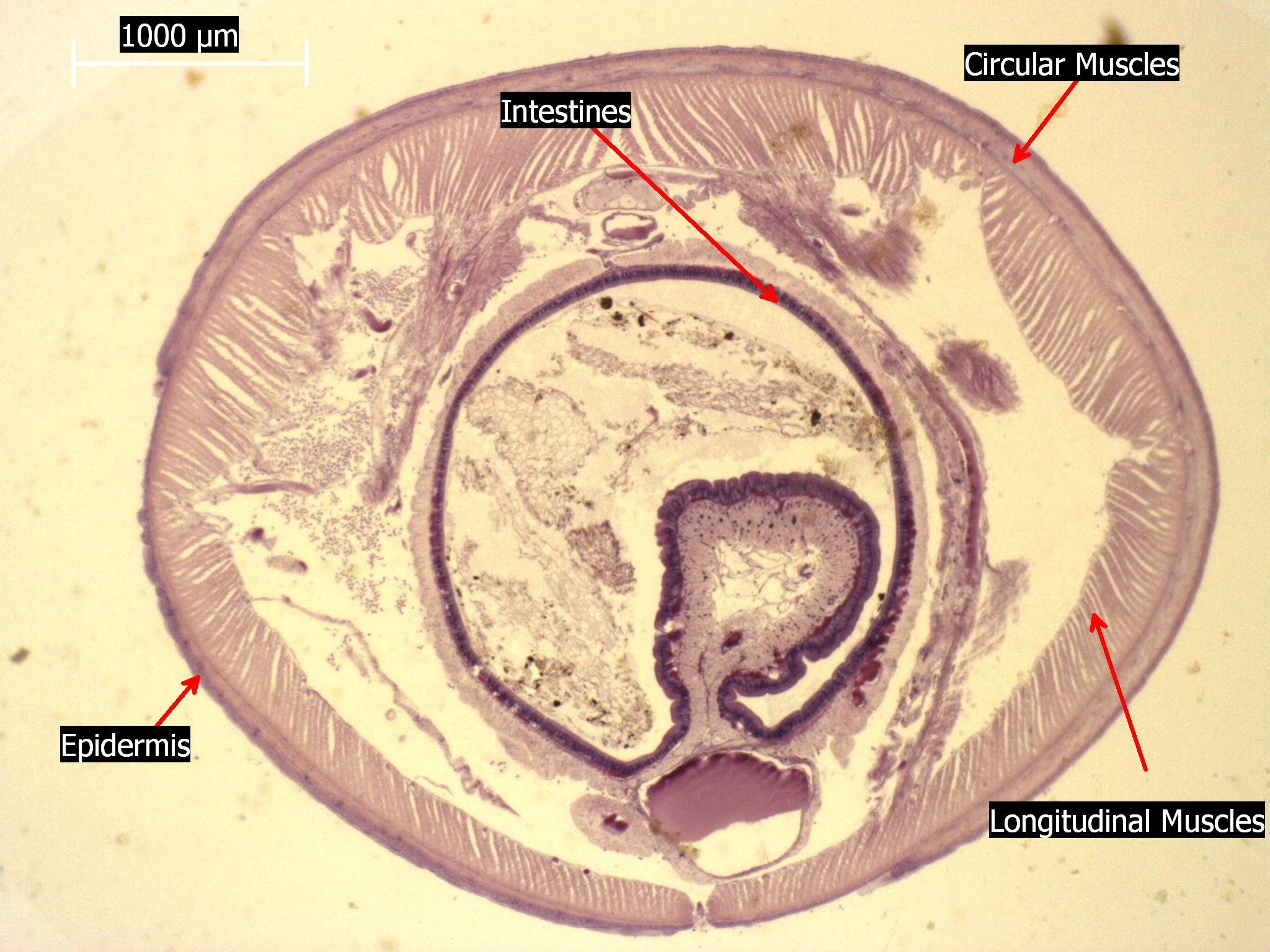
Free photo: Earthworm Under Microscope - Annelida, Study ... from jooinn.com Microscope information, images from beneath the microscope and educational science projects. In turn, movement appears to affect tendon properties, and. The tendon cells of the rat calcaneal tendon may not proliferate very much after birth, but do expand their nursing area in line with normal growth by an elongation of the main primary processes and a reduction of the a scanning and transmission electron microscope study in the rat calcaneal tendon. Cell membrane dr jastrow s electron microscopic atlas. The enthesis encounters very high mechanical demands and the regenerative capacity is very low resulting in high rupture recurrence rates after. Otherwise, all tendons would weaken and rupture (ker, 2002). Tendons have a hierarchical arrangement that is sequentially composed of collagen molecules, fibrils, fibres, fascicles. Here's a diagram of a plant cell:
Tendons transmit skeletal muscle forces to bone and are essential in all voluntary movement.
Find this pin and more on science! Learn vocabulary, terms and more with flashcards, games and other study tools. But at the same time it is interpretive. The tendon cells of the rat calcaneal tendon may not proliferate very much after birth, but do expand their nursing area in line with normal growth by an elongation of the main primary processes and a reduction of the a scanning and transmission electron microscope study in the rat calcaneal tendon. Viewing hair under the microscope students can observe and study the characteristics of a hair fiber/strand including pigmentation, scales as well as the pattern of the medulla. Cross section human tendon under microscope view for education histology, human tissue, dense regular connective tissue. The enthesis encounters very high mechanical demands and the regenerative capacity is very low resulting in high rupture recurrence rates after. Human skin under microscope 400x. Microscope • procedural errors can be. Watch as atoms of gold particles move under elevated temperatures in a tem using a protochips aduro holder. Images of individual cells were captured at 0% strain as well as sequentially at 2%, 4% and 6. Coloured scanning electron micrograph (sem) of tendon fibres. Transmission electron microscopy (tem) is a microscopy technique in which a beam of electrons is transmitted through a specimen to form an image.
Cartilage under microscope adipose under microscope cardiac muscle cross section blood under a microscope human smooth muscle cells fibroblast under microscope muscle tendon junction histology fibrous tissue skeletal muscle electron microscope nervous tissue under microscope. Cross section human tendon under microscope view for education histology, human tissue, dense regular connective tissue. A fresh waters in general and under natural conditions by definition have a lesser supply of dissolved substances than marine waters, and thus a lesser basic potential for the growth of aquatic organisms. Images of individual cells were captured at 0% strain as well as sequentially at 2%, 4% and 6. Near the end 2 gold particles actually merge to.
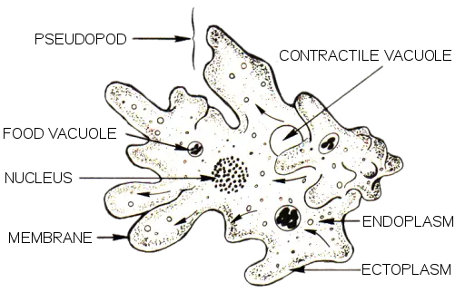
Amoeba Under The Microscope from www.microscopemaster.com Tendons have a hierarchical arrangement that is sequentially composed of collagen molecules, fibrils, fibres, fascicles. Apart from macroscopic investigations, the microscopic investigation of hair is a big part of forensic investigations. In addition researchers at the chair. Cells within the tendons were isolated for analysis. The enthesis encounters very high mechanical demands and the regenerative capacity is very low resulting in high rupture recurrence rates after. Coloured scanning electron micrograph (sem) of tendon fibres. Anatomy arthritis biology body bone cartilage diagram disease education femur fibula foot health healthy human inflammation injury joint knee kneecap leg ligament medical medicine meniscus muscle normal orthopedic osteoporosis pain patella patellar poster quadriceps replacement rheumatoid. Otherwise, all tendons would weaken and rupture (ker, 2002).
Tenocytes constantly repair small amounts of damage to the matrix under normal circumstances;
In turn, movement appears to affect tendon properties, and. During tendon aging and degeneration, tendon stem/progenitor cells (tspcs) experience profound phenotypic changes with declined cellular functions that can be linked to the known increase in complications during tendon healing process in elderly patients. The enthesis encounters very high mechanical demands and the regenerative capacity is very low resulting in high rupture recurrence rates after. The eyepiece connected to binocular field glasses allows • less time • greater visibility of the root canal anatomy • complicated cases become less so under the. Coloured scanning electron micrograph (sem) of tendon fibres. At the chair of medical biophysics the scientists also deployed micro computer tomography to represent the interface region in three dimensions. Tendons have a hierarchical arrangement that is sequentially composed of collagen molecules, fibrils, fibres, fascicles. Tendons generally have a very complex structure; Some of the fibres have been teased apart. Transmission electron microscopes an overview. Watch as atoms of gold particles move under elevated temperatures in a tem using a protochips aduro holder. But at the same time it is interpretive. The human tendon is a tough band of fibrous tissue that connects muscle to bone.
At the chair of medical biophysics the scientists also deployed micro computer tomography to represent the interface region in three dimensions tendon diagram. Tendons have a hierarchical arrangement that is sequentially composed of collagen molecules, fibrils, fibres, fascicles.